
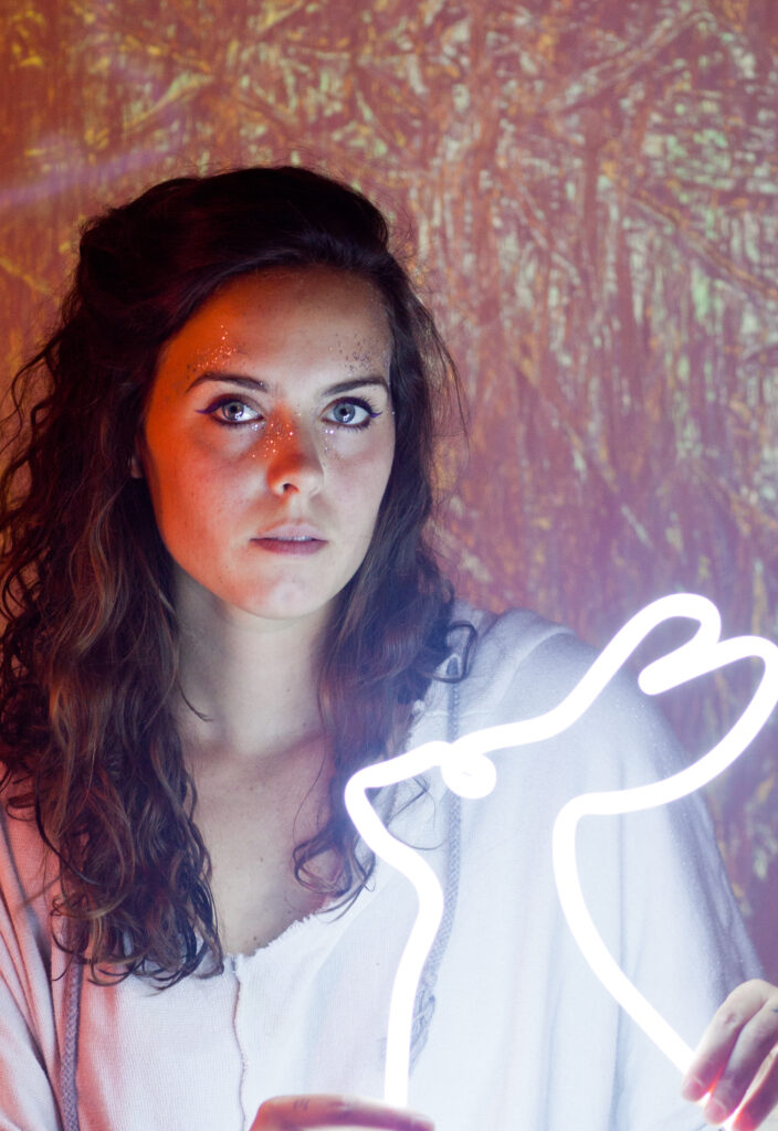
Delivering complex information to the masses is not easy. Artists have always stretched the boundaries of concept and creativity. The goal of our Artist + Researcher (ARx1) Exhibition is to see what happens when the talents of both science and art join forces to conceptualize a way to communicate the complex.
The Phoenix Bioscience Core established the Artists + Researcher Program with Phoenix-based Bentley Gallery to connect the complex life science research happening on the PBC with a wider audience. Nine researchers from each of the three public universities — Arizona State University, Northern Arizona University and the University of Arizona — worked alongside local artists to establish a transformational piece of artwork that shares their research in a new medium.
All of these works of art + science will then come together to form the first Artist + Researcher Exhibition Tour. The Artist + Researcher Exhibition is one of many programs within the Art in Medicine initiative on the Phoenix Bioscience Core. The initiative aims to explore what happens when research is presented through an artistic lens.
The first cohort of the PBC Artists & Researchers program finished their projects in the spring of 2022. They will be on display around Arizona during the 2022-2023 academic year. The first public display will be on September 2nd at the Health Sciences Education Building.
Explore the work
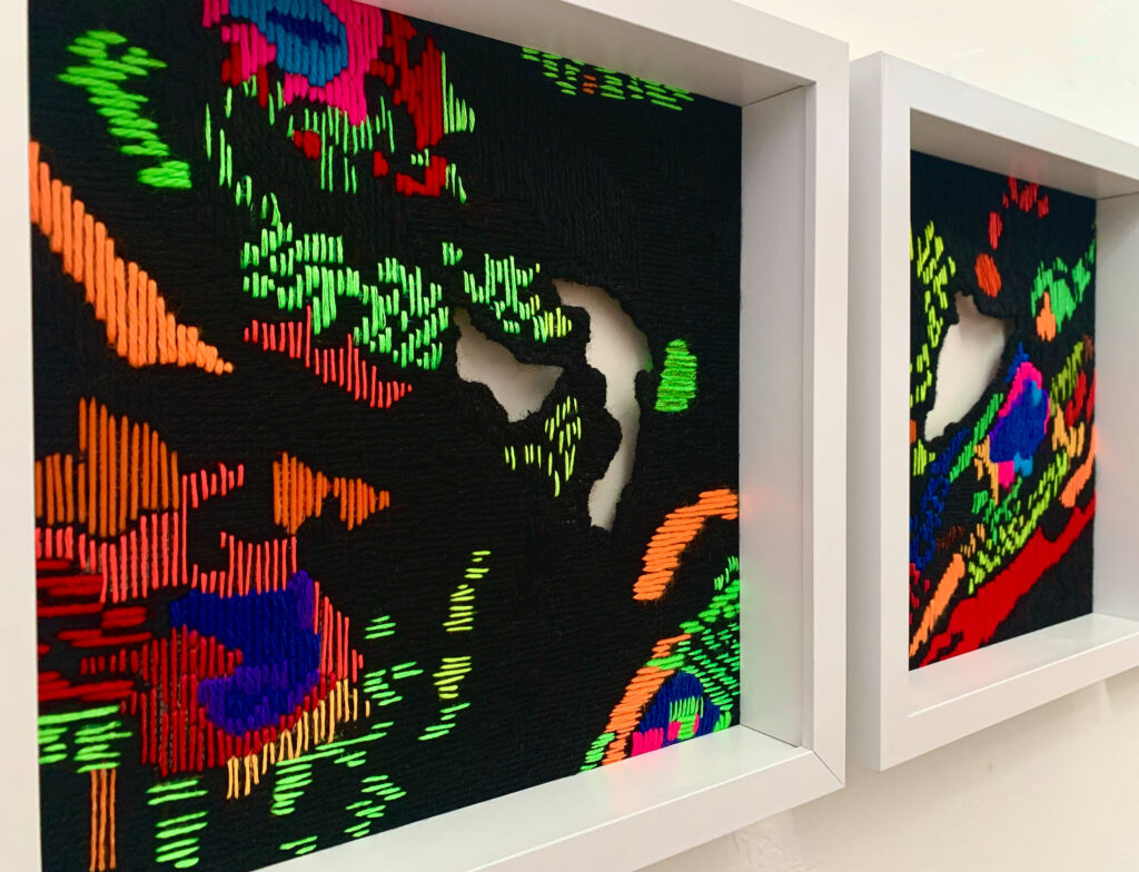
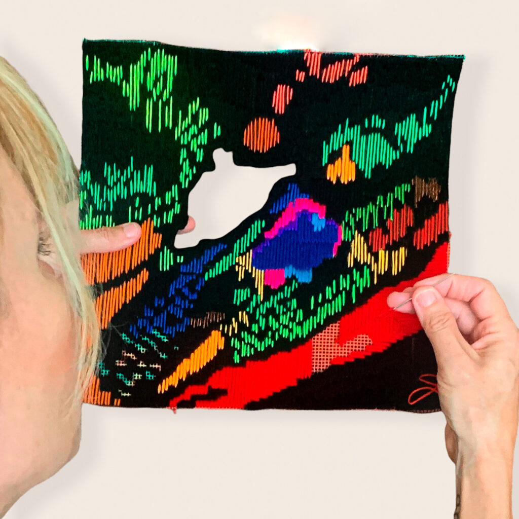
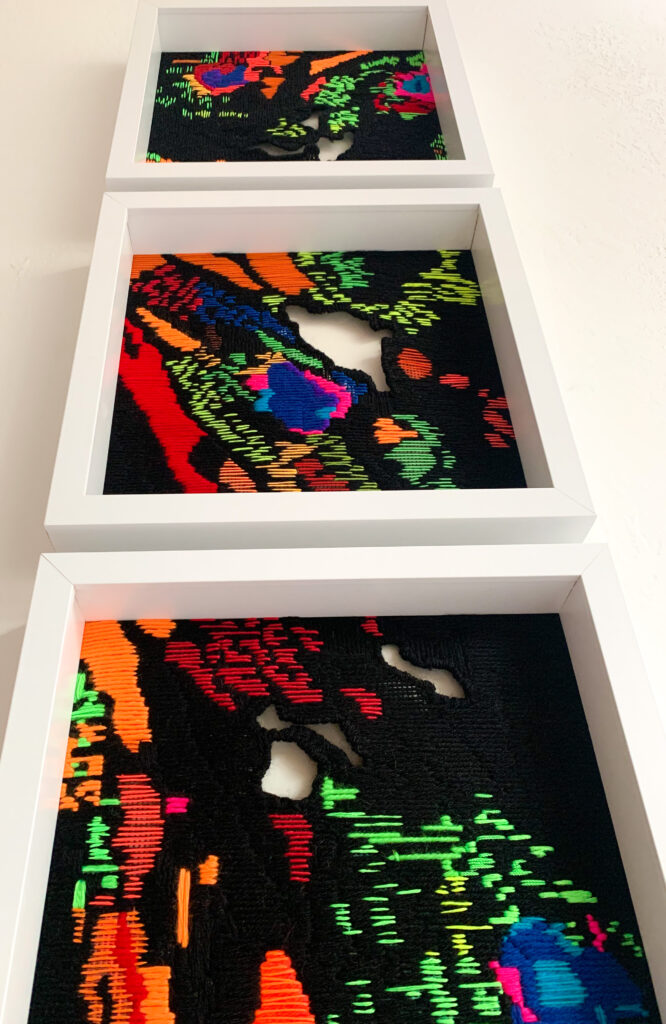
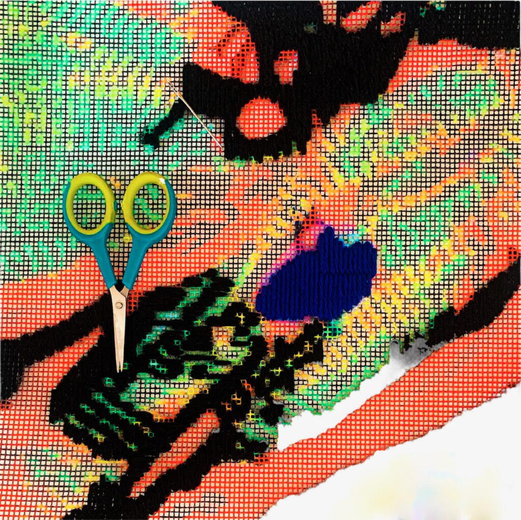
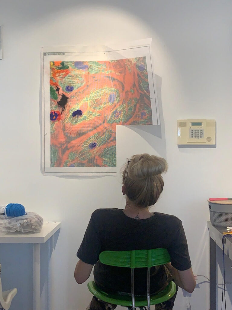
Layers of Cardiac Fibers
Denise Yaghmourian + Dr. Tobias Jakobi
During the process of collaboration, we both found the stained heart muscle imagery very interesting. We decided that a few particular images were very inspiring and also aesthetically interesting.
Below describes visual imagery used for the collaborative process.
Tobias Jakobi:
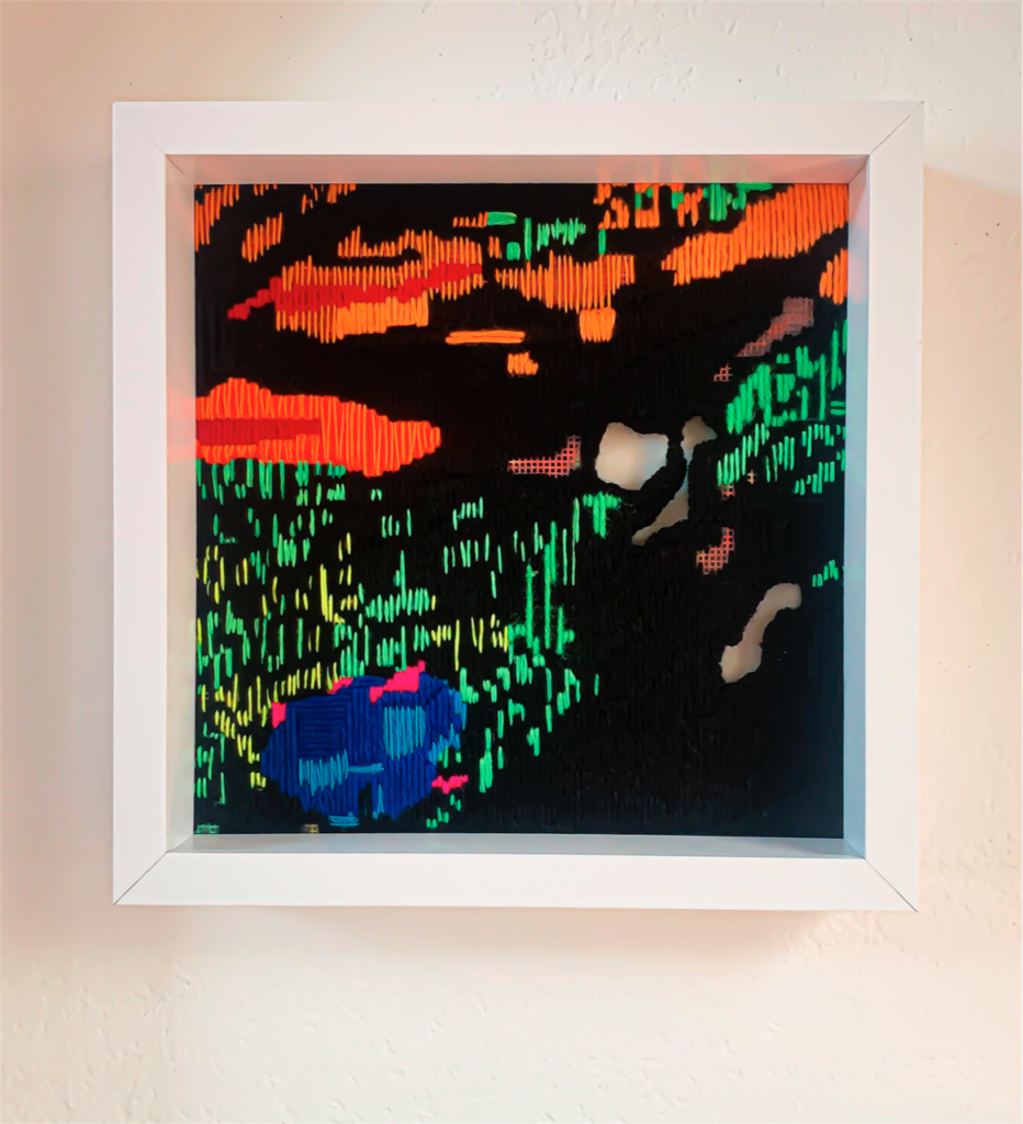
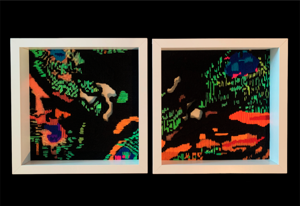
In my lab, we aim to understand cardiovascular disease at the level of RNA, which cells usually use as a blueprint to make proteins. Some RNAs however, do not act as blueprint and seem to have other purposes. These molecules are specifically interesting to my lab as they might regulate important cell functions. To investigate those functions, we often stain heart muscle cells using fluorescent dyes, which are visible under the microscope using specific wavelengths of light to make each of the colors emit light that in the microscope is translated into a distinct color. These photos are actually a merge of three different images using the three different colors since we cannot see all colors at the same time under the microscope.
Blue is often used for the cell nuclei (where the cell’s genome is stored). In green we stained a protein that is located in the muscle fibers that run throughout the cell. Since this protein is part of specific structures called disks in the muscle fibers, we see a characteristic pattern in green. The red color is used to visualize a protein of the cell’s internal structure, also called the cytoskeleton, of cardiac muscle cells.
Denise Yaghmourian:
I am particularly fascinated with these images as a way to show and observe the heart. I worked experimentally with fibers and threads in an organic way to create these 3 pieces. Each of the 3 pieces pay particular focus to highlighting the fluorescent dyes, visible under the microscope. The pieces were created one small stitch at a time on mesh material. During the process I thought a lot about the connectivity of the fibers of heart muscle cells and the amazing ways they all work together. I was also interested in experimenting with using traditional needlepoint practices in a modern way. The separate stitch is representative of heart muscle cells, with focus on the fluorescent dyes as visual imagery. I created open areas amidst the threads as a way to feel dimensionality and intimate connection to the human heart.
The Artist: Denise Yaghmourian
Denise Yaghmourian is a mixed media and installation artist who works with a variety of materials, including paper pulp, paint, thread, fabric, vinyl and found objects. Her works are hybridized forms, with methodological roots in past movements like Minimalism and Post-Minimalism, and a conceptual grounding in contemporary practices. The simplicity of her surfaces reveal a material-based sensibility. Conjoining the repetitive process inherent in machine-made items with laboriously repetitious handwork, Yaghmourian embraces the hypnotic and the meditative.
The Researcher: Dr. Tobias Jakobi

Assistant Professor, Department of Internal Medicine and a member of the Translational Cardiovascular Research Center (TCRC) at The UA College of Medicine – Phoenix. As a bioinformatician, his research interests center on the interconnection of wet lab research and computational research. His lab focuses on the interplay of different RNA species and their underlying functional networks in the heart, as well as developing new computational open source tools that can be used by other researchers in their field of interest.
More Information
Medium: Assorted embroidery threads, yarn, paint on mesh
Retail cost: $750 each
‘It’s All In Your Head’ – Deconstructing the brain after concussion
Rembrandt Quiballo + Dr. Theresa Currier Thomas
Often, the persisting symptoms of concussion are dismissed with the statement ‘It’s all in your head.’
Over 55 million concussions are reported to hospitals a year and more than that are never reported. Over 50% of concussed individuals experience ongoing cognitive decline, mood disorders, chronic headaches, sensory deficits, hormonal dysregulation, sexual dysfunction, and changes in sleep patterns. Concussed individuals are at higher risk for early-onset neurodegenerative diseases, cerebrovascular events, and cardiovascular disease. The social effects are profound, disintegrating interpersonal relationships and ability to hold a job. Despite having severe life-altering effects, ‘we’ can’t see the damage, the symptoms are generic, and concussed individuals become invisible, ignored and dismissed by family and healthcare providers, never receiving the medical and social support needed to help compensate for damage and persisting changes going on in their brain.
Using Augmented Reality technology, the viewer can walk through the brain, entering different compartments where specific cell types are highlighted and pathology can be detected by the naked eye. Using an experimental model of a moderate-severe concussion, sections of the brain have been reconstructed in 3D, with compartments containing normal (left side) and concussed brains (right side) demonstrating cells and structures mediating neuroinflammation (microglia), homeostasis and blood-brain barrier permeability (astrocytes), neuropathology and vasculature (silver stain), white matter tracts (myelin stain), and general cellular distribution (nuclear stain).
Interactive compartments allow for compare and contrast the impact of brain injury by enlarging sections to compare the pathology and regional distribution of damage. Visualize the damage that has so long been invisible.
With this perspective, the ambiguous statement, ‘it’s all in your head,’ is no longer a dismissal, but the actual reality.
More information
Dr. Currier Thomas studies the longitudinal changes in the brain that underlie persisting and developing post-concussive symptoms to identify structural, molecular, and functional changes relevant to specific symptoms (sensory, cognitive, emotional). Growing up with a family member who suffered a mild TBI, she is overly aware of the negative stigma associated with symptoms, the profound impact on family dynamic, and the neglect of proper education and support. Her research demonstrates persisting, dynamic, brain pathologies, sex-differences, and functional differences. Her interactions with Rembrandt have created an effective educational tool that tells a story of the brain’s complex response to concussion and provides profound visual evidence capable of promoting understanding/acceptance, increased patient and family support, and the overwhelming need for continued research.
Rembrandt is an adept and patient listener and learner, asking critical questions and taking the time to understand my passion for brain injury research. At first, I couldn’t see it – but as the project has evolved, he nailed it, he got me. He’s created the ultimate artwork that can help communicate to the general population the profound impact of concussion on the brain, the ultimate reference to demonstrate what goes wrong, so we, as a community, can better educate, provide better health care, and better describe the rational for our research and the impact. Brilliant.
The Artist: Rembrandt Quiballo
Soon every facet of human life will be digitized. My work imagines the possible remnants of our existing digital culture. While archaeologists excavate the earth to find physical evidence of long lost civilizations, my work shows the possibilities of what future scientists would discover in our ever expanding digital cloud.
Through the use of glitch techniques I generate digital artifacts within images found in mass media such as film, television and the Internet. The prevalence of data compression today makes this loss of information inherent in our everyday images. We want unsurpassed quality but with the least expenditure of resources. The mere transfer of data causes the contemporary image to be in a constant state of decay. In our pursuit to produce and consume an endless stream of visuals, we face complications such as finite data space and visual incongruity. In the meantime, a new kind of imagery emerges.
Quiballo was born in the city of Manila in the Philippines. Social and political unrest in the Philippines would compel his family to leave the country, eventually immigrating to the United States. Quiballo received a BFA in Painting and a BA in Philosophy from the University of Arizona and holds a MFA in Photography from Arizona State University. His works have been exhibited nationally and internationally including The Lab, San Francisco, Phoenix Art Museum, Phoenix, Medrar for Contemporary Art, Cairo, Labor Neunzehn, Berlin and Durden and Ray, Los Angeles
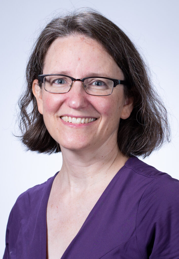
The Researcher: Dr. Theresa Currier Thomas
Dr. Currier Thomas’s primary research interests focus on traumatic brain injury (TBI). She studies structural, functional and molecular processes, with synaptogenesis as a keystone, which guide circuit reorganization over time and contribute to chronic deficits/symptoms after TBI. She tests pharmacological and rehabilitative strategies to mitigate these chronic deficits. She is also actively investigating the contribution of endocrine (hormone) deficiencies after TBI to understand causes and propose treatments for post-traumatic neurological deficits. These studies are in collaboration with researchers from Barrow Neurological Institute at Phoenix Children’s Hospital, University of Arizona College of Medicine – Phoenix, Phoenix VA Healthcare System and Arizona State University.
More Information
Medium: Augmented Reality Installation
Retail cost: $10,000
Deciphering The Nature of Cardiokines
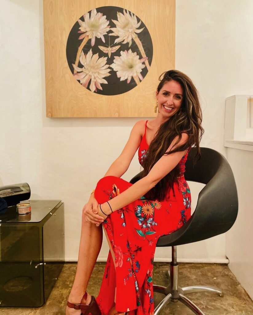
Alexandra Bowers + Dr. Shirin Doroudgar
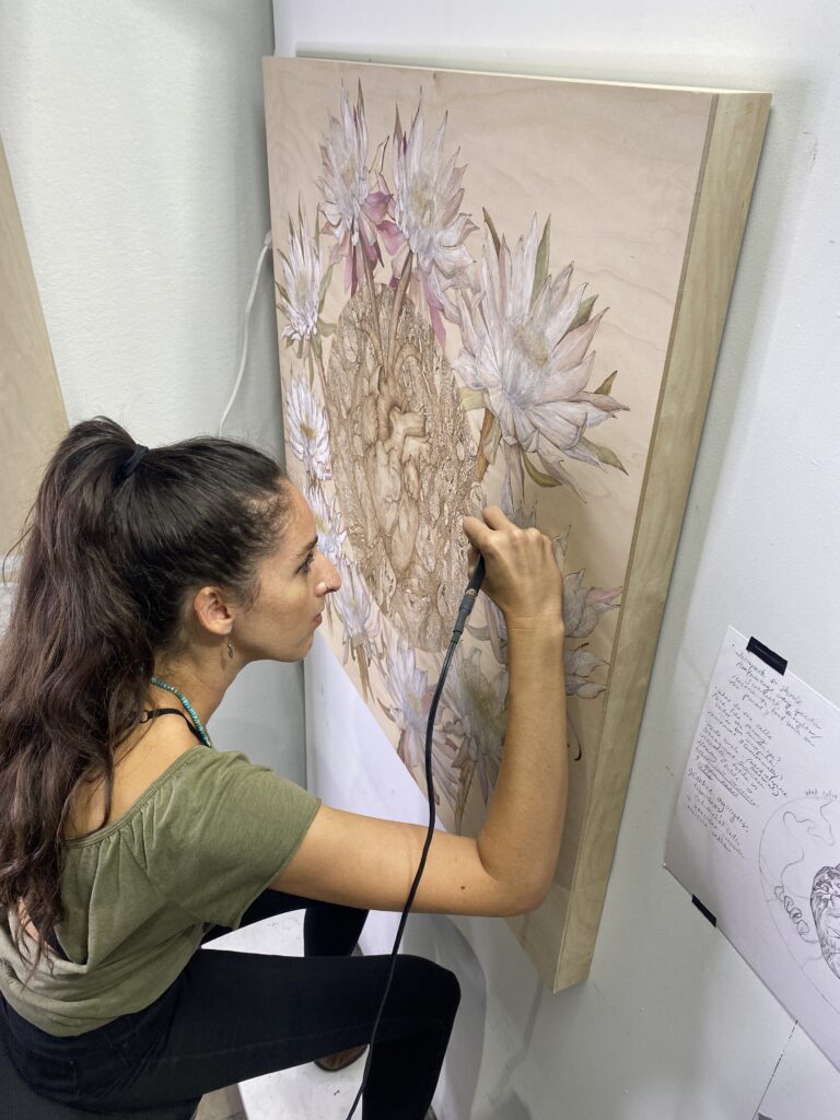
“Deciphering the nature of Cardiokines” is a symbolic and literal depiction of communication between the heart cells Dr. Shirin analyzes under her microscope, and a specific flower species “Peniocereus Greggii” also commonly referred to as “Queen of the Night” Alexandra highlights in her artwork. Part of Dr. Shirin’s lab work is understanding cellular communication systems that begin in the heart and then interact with the rest of the body. One of the reasons I chose to depict the “Queen” is because of a certain communication network that allows for synchronistic blooming for one night each year. One of the key themes of this partnership was learning a new language from both the perspectives of the researcher and the artist. The dialogue we shared throughout this process was meaningful and heartfelt. “Deciphering the Nature of Cardiokines” is meant to highlight two unique realities that intersect because of conversation.
More information:
Alexandra Bowers is a Phoenix based pyrography/mixed media Artist. Dr. Shirin Doroudgar is an Assistant Professor of Internal Medicine at the Translational Cardiovascular Research Center at University of Arizona. “Deciphering the Nature of Cardiokines” is about the intersection between Dr. Shirin’s heart research and Alexandra Bowers’ love of nature. The flower species, Peniocereus Greggii, was specifically chosen because of the communication system inherent in the genetic makeup of the flower. The myocardial tissue around the heart was depicted to convey the imagery of what Dr. Shirin observes from behind her microscope in the lab. Throughout the process of this partnership, I was most inspired by the concepts of learning the scientific vernacular that Dr. Shirin uses within her field, as well as the effortless communication that we had with one another. Above all, I value the development of a new friendship with someone I most likely would have never crossed paths with had I not been invited for this opportunity. The primary intention of this art piece is to emphasize the concept of conversation from both a literal and figurative perspective, simultaneously linking each passion that both artist and researcher have for the work they’ve dedicated their lives to pursuing.
The Artist: Alexandra Bowers
My name is Alexandra Bowers, and I was born and raised in the Sonoran Desert. As an artist focusing on the natural world, my intention is to find ways the environment and our personal lives intersect and converse with one another. Utilizing the wood burning process technically known as pyrography, allows me to illustrate with heat the indigenous plants and animals that live within our neighborhoods and urban streets. Over the years I’ve witnessed technology and made-made structures detach us physically and psychologically from our arid surroundings. Urban sprawl has forced our habitat to either vanish or move away. As we pave, develop, and grow our urban cities, the natural environment diminishes and is forced to change and adapt to its human neighbors. My work is concerned with highlighting these beings, to bring our awareness and emotional empathy back to them before they move on, or permanently disappear.
The Researcher: Dr. Shirin Doroudgar
Shirin Doroudgar, PhD is Assistant Professor of Internal Medicine at The University of Arizona College of Medicine – Phoenix and the Translational Cardiovascular Research Center (TCRC). Her research program utilizes integrative approaches from molecular and organellar to cellular and organismal levels to examine the dynamics of the protein homeostasis network in health and disease. The Doroudgar Lab’s current focus is on studies of cellular stress responses that underlie the dynamic remodeling of the cardiac myocyte proteome and secretome.
More Information
Medium: Pyrography (Wood Burning) and Water Soluble Wax Pigment
Retail cost: $10,000
Invisible War
Bill Dambrova, Dr. Cynthia Ivy + Dr. Gretchen Bachman
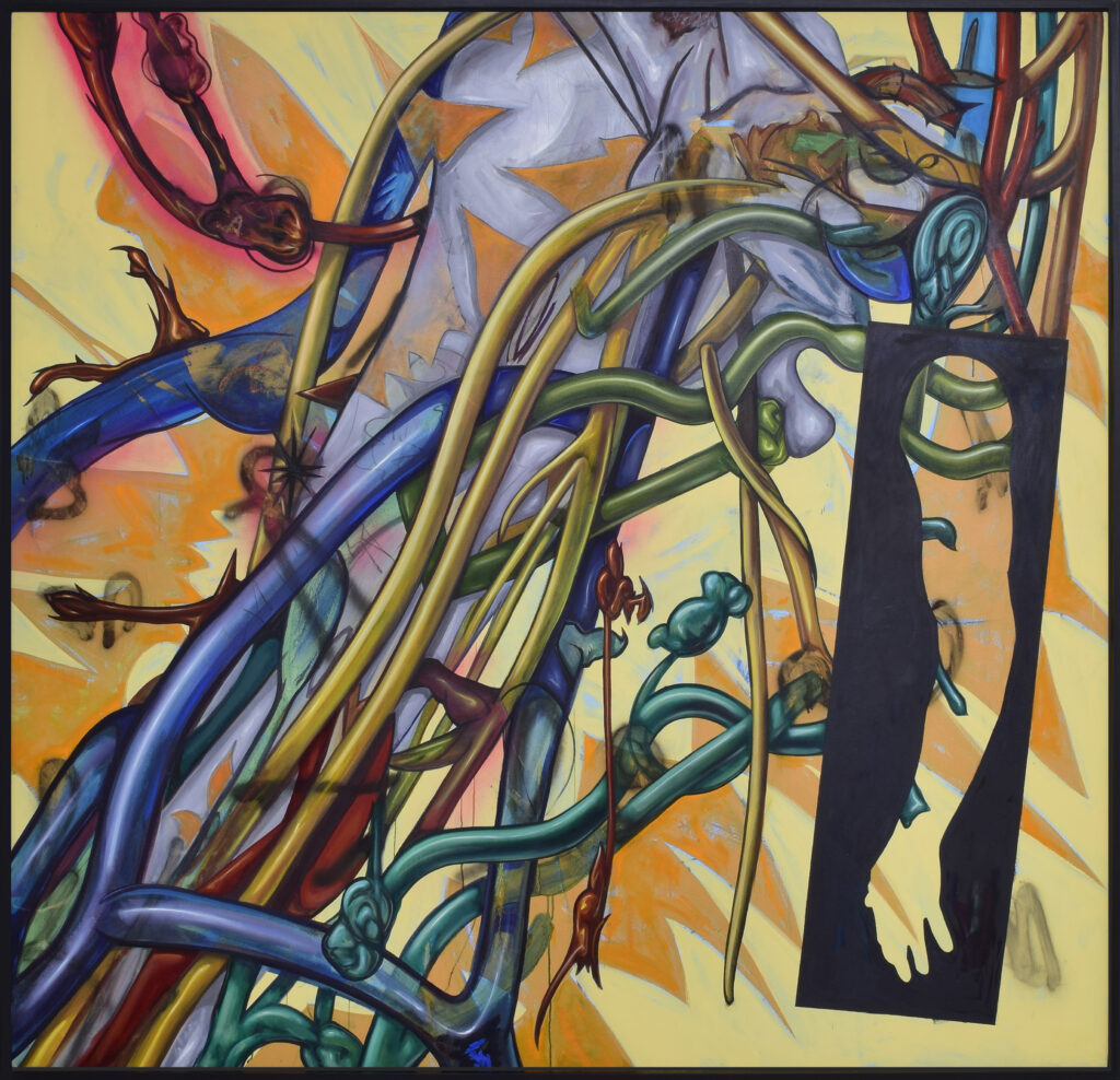
Art can stimulate questions and inspire new perspectives that spark epiphanies and solutions to problems. From the start, we knew we wanted to avoid making a medical illustration that attempted to describe the experience of Complex Regional Pain Syndrome (CRPS) for any given individual. We wanted the work to be about feeling and empathy for the person who has CRPS, so as a team, we decided that an abstract or stylized painting would be the best representation. The approach was for Bill to process and visualize the information as Cynthia and Gretchen explained the disease and their research. Then when beginning the work, he allowed the imagery he was getting in his head when they described it to unfold from his subconscious as he created the initial expressive marks on the canvas. The first marks that Bill made are not seen in the finished work, but the energy released while making them set up the back and forth interactions on the canvas that emulate what he imagined to be a constant ongoing battle in a person’s body while they are fighting the disease. This was a very fast energetic process with the intention of exuding energy, frustration, and pain that a person with CRPS may experience. At some unnamable point in the process of creating the abstract marks, a story of sorts started to form for Bill and he began to push the marks in that direction.
From the artist:
Because the presentation of CRPS and individual experiences with CRPS are variable, we want to remind the viewer that that this painting is in no way an actual representation of CRPS. No two people experience this syndrome identically and none of us on this team have ever been diagnosed with CRPS.
Our team’s goal with this project is to create a monumental, beautiful, and powerful piece of artwork that acknowledges the invisible battles in the war between the person and their disease. The intention is to acknowledge the experiences of those diagnosed with CRPS and increase awareness to the general public. We encourage and support further inquiry and research to destigmatize CRPS through art and education.
The Artist: Bill Dambrova
Bill Dambrova is a native Arizonan and ASU graduate with a BA in Studio Art. Bill has shown artwork in local as well as national galleries and museums, and has created site specific art installations at locations including: Valley Metro Rail, Burning Man, East Jesus, and Meow Wolf. His work can be found in the public collections of institutions such as The Tucson Museum of Art, Sky Harbor Airport Museum, The City of Scottsdale, The State of New Mexico: Art in Public Places Program, DuPont, Kroger, Kemin Industries, and The Phoenix Suns Legacy Partners.
Bill is moving into new territory as a Public Artist. He has completed the fabrication phase of a 6000 square foot terrazzo floor he designed for the newly built Sky Harbor Airport Rental Car Return Train Station in Phoenix, Arizona. The station is scheduled to open to the public September 2022.
Bill’s next solo museum exhibition of large-scale paintings will be at the Mesa Contemporary Art Museum in Mesa, AZ opening in 2023.
The Researcher: Dr. Cindy Ivy

Cindy Ivy is a clinical professor of Occupational Therapy at Northern Arizona University (NAU) at the Phoenix Bioscience Core. Her research has focused on interventional research for hand and upper extremity and Parkinson’s Disease. She teaches anatomy to occupational therapy, physical therapy and physician assistant students at NAU and University of Arizona medical students. She also teaches a number of other classes including a course on the upper extremity with Dr. Bachman. She is a certified hand therapist. In addition, she enjoys spending time with her four adult children, Jacob, Caroline, Calia and Jonah, creating pottery and other forms of artwork, and enjoying the arts and the outdoors.
Collaborative Intervention Research Interests: Complex Regional Pain Syndrome. Quality of life or studying quality of life measures in disabilities, wearable intervention, biomechanical studies, anatomic studies, ALS/Motor Neuron Disease, Parkinson’s Disease and exercise science.
The Researcher: Dr. Gretchen Bachman
Gretchen Bachman is an Assistant Clinical Professor for the Occupational Therapy Program at Northern Arizona University, located at the Phoenix Bioscience Core. She teaches a variety of courses in the program related to leadership and management, physical disabilities, evaluation and outcome measures, scholarship, environmental adaptation & assistive technologies, and courses on hand/upper extremity injuries and disease. Gretchen is a Certified Hand Therapist and serves in many roles associated with national hand therapy and surgery organizations. She travels annually with the Guatemala Healing Hands Foundation, an organization dedicated to improving the quality and availability of healthcare in Guatemala through education, surgery, therapy, and community development.
Collaborative Research Interests: Complex Regional Pain Syndrome, sleep, quality of life, health equity, and upper extremity function and dysfunction.
More Information
Medium: Mixed media on canvas, Framed in black 74” x 74”
Retail cost: $14,000
Tissue Study I
Mark Pomilio + Dr. Taben Hale
After agreeing on a date and time, I visited Dr Hale’s research lab and was introduced to her work. During our meeting, Dr Hale outlined her methods and goals through discussion and examples of which revealed the materials and systems utilized to achieve the desired outcome. Aspects of her research involve documentation through digital imaging. These images are both informative and visually fascinating. I asked Dr Hale to send several of these images, and they became a starting point for my inquiry.
Description of the piece
Tissue Study #1 represents an integration of my creative mathematical, geometric methods developed over the last several decades and those images I experienced through the visual recordings of Dr Hale’s research with cardiac tissue pathological remodeling. First, I began by triangulating the relationships between two geometric forms: the pentagon and hexagon. These two forms are often utilized when expressing chemical cell structures in scientific documentation. During each painting session, I added an additional layer of geometric forms, which were connected to the previous layer of forms through additional triangulation. This method was my attempt to mirror the naturally occurring building systems found in the myocardial connective tissue which provides the structural scaffolding for the heart cells and thus, relating my structural methods to that which I experienced in Dr Hale’s research imagery. This building method extended into many painting sessions and ultimately represents upwards of nearly twenty layers, one applied to another, with subtractive and additive decisions made throughout the long painting process.
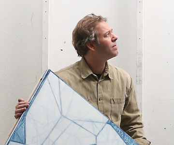
The Artist: Mark Pomilio
Mark Pomilio’s current research has focused on creating images, which embody principles of geometry, fractals, cloning and single cell manipulation. These interests have led to invitations to lecture nationally on topics as diverse as art and mathematics and the role the visual arts can play, in understanding the social ramifications of advances in the Life Sciences. His artwork has been featured in solo museum and gallery exhibitions nationally and internationally, including Luxehill Museum in Chengdu, China, Xu Beihong Art Academy, Renmin University of China, in Beijing, the Chapelle Saint-Louis de la Salapetriere, in Paris, France, and Art Resources Transfer, in New York City.
Originally from Philadelphia, Mr. Pomilio is currently living and working in Phoenix, Arizona. Where he is a Professor of Painting and Drawing within the School of Art, at Arizona State University’s Herberger Institute for Design and the Arts.
The Researcher: Dr. Taben Hale
Dr. Taben Hale is an Associate Professor in the Department of Basic Medical Sciences and Director of Women in Medicine and Science at the University of Arizona, College of Medicine – Phoenix. She joined the University of Arizona, College of Medicine – Phoenix in 2008 as one of the founding faculty in the Department of Basic Medical Sciences where she leads a research program and teaches pharmacology to 1st and 2nd year medical students.
Dr. Hale’s research, which has been funded by the National Institutes of Health and the American Heart association, focuses on the causes and consequences of high blood pressure. Specifically, her team investigates the molecular and cellular mechanisms of pathological cardiac remodeling that are crucial to our understanding and mitigation of hypertensive heart disease. In addition, her team researches the early drivers of cardiovascular disease that may be rooted in the in utero environment.
More Information
Medium: Oil on linen
Retail cost: $8,000
Long Division # 7
Michael Marlowe + Dr. Jonathan Lifshitz
Biomedical science is an art. Both science and art are systematic processes. The process includes a mission, a plan, trial, error, implementation, and analysis. The pieces created represent the scientific and artistic process. When the finished piece appears straightforward, it is because substantial thought, effort, and time went into creating it.
The pieces represent the execution of a clear plan, knowing that variance, error, and wobble will shine through. Like mowing an expanse of grass, the process appears simple, until the lines deviate, the rows overlap varying amounts, and the corners add a new challenge. In the pieces, from a distance, the systematic plan is clear. In the details, the wobble through the lines becomes evident. The inconsistent pace of the brush becomes evident. The variance in the pigment becomes evident. And the analysis of the final product reflects the lanes, bands, and staining achieved in standard protein electrophoresis gels known as western bolts.
In the end, the parallel lines of varying thicknesses and densities also represent our multiple marginalized identities as scientists and artists. In some respects, the lines mirror the multitude of gender identities and expressions traversing the array of PRIDE flags.
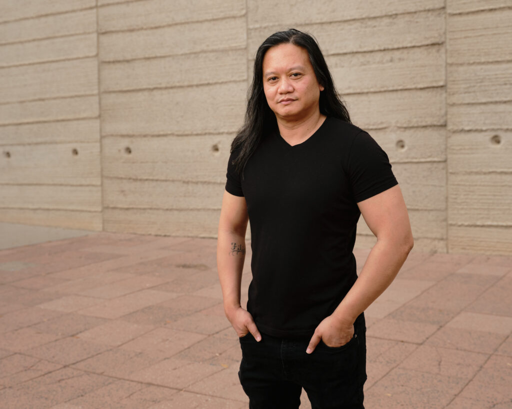
The Artist: Michael Marlowe
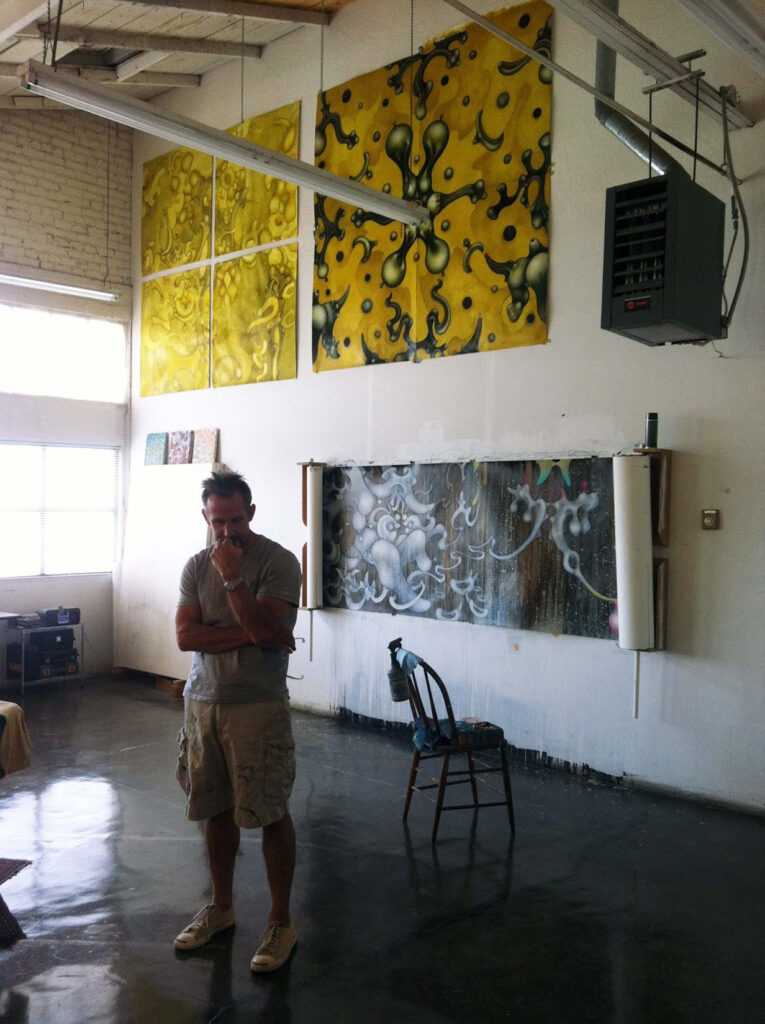
Michael Marlowe is a studio artist, art director and production designer working in the film and television industry. Marlowe’s large-scale painting process takes an abstract approach to imagery that is rooted in figurative representation. His works examine and vibrantly animate aspects of the human figure from the inside out. Interested in various modes of self exploration, he visually abstracts and reshapes his subject in a pursuit to visualize the human form as something expanded, exploded, and restructured. “It’s the aspects of self we don’t show, the aspects of self we very possibly don’t know that make us tick. This visual deconstruction, this opening up, is in order to see around the parts we know, to discover the parts we don’t.”
The Researcher: Dr. Jonathan Lifshitz
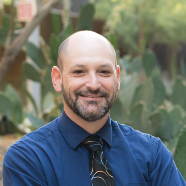
Dr. Lifshitz is a neuroscientist with specific subject matter expertise in traumatic brain injury. He is a Research Professor at the University of Arizona College of Medicine-Phoenix. He has a passion for creative studies to understand how the brain injury dismantles, repairs, and regenerates circuits in the brain, with a focus on inflammation and rehabilitation. He takes immense pride in mentoring and training others in the scientific processes that generate evidence to inform clinical decisions. In the community, he raises awareness and develops services for traumatic brain injury in the context of domestic violence.
More Information
Medium: hand-painted watercolor on cold pressed Arches paper
Retail cost: $8,800
Dyad
Lily Reeves + Dr. Shelby Langer
Dyad is inspired by Dr. Langer’s research on dyadic or two-person communication in the context of health and illness. This work has spanned multiple dyad types, including persons with advanced cancer and their caregiving partners, children with chronic pain and their parents, and patients and their healthcare providers. Much of this work has found that behaviors intended to buffer the other (e.g., hiding concerns or being overprotective) are ironically associated with increased distress.
The two mirrors use color, perspective, and optical illusion to speak to the combination of two truths. Effective communication requires not just open sharing of thoughts and concerns but also responsive listening such that the discloser feels truly understood and validated. By standing in the center of the piece, and relaxing your eyes, a third, phantom image appears at the center of the installation. If there are others standing in front of the two body-sized mirrors, you see a combination of these two portraits within the third phantom light image. This purely psychological mirage is a physical depiction of the act of communicating and understanding. Mirrors have long been a visual representation of the self, and by default, the outside-the-self. This piece gives us a moment to ask ourselves: How can we learn to care for others? How can we learn to incorporate others’ truths into our own world view, to cultivate a world that is more understanding? How do we create space for listening to each other? For honest conversations? For understanding something other than ourselves?
The Artist: Lily Reeves
Reeves’ practice is multifaceted, but is always grounded in the medium of light and energy. By using light, space, collaboration, and immersive environments, Reeves strives to make room for openness and magic in a world that is increasingly disenchanted. Reeves’ conceptual work explores the emotional, psychological, and spiritual worlds of personal, societal, and environmental healing.
Lily Reeves is originally from Birmingham, Alabama. She holds a Bachelor and Masters in Fine Art from Alfred University and Arizona State University. Reeves helps facilitate an artist residency and arts-related programming for the Takoja Institute, a center for creative and spiritual healing, in Taos County, New Mexico, where she serves as acting Artist Coordinator. Reeves currently lives in Phoenix, Arizona and runs her Art and Design Studio, Reeves Studios, full time.
The Researcher: Dr. Shelby Langer
Reeves’ practice is multifaceted, but is always grounded in the medium of light and energy. By using light, space, collaboration, and immersive environments, Reeves strives to make room for openness and magic in a world that is increasingly disenchanted. Reeves’ conceptual work explores the emotional, psychological, and spiritual worlds of personal, societal, and environmental healing.
Lily Reeves is originally from Birmingham, Alabama. She holds a Bachelor and Masters in Fine Art from Alfred University and Arizona State University. Reeves helps facilitate an artist residency and arts-related programming for the Takoja Institute, a center for creative and spiritual healing, in Taos County, New Mexico, where she serves as acting Artist Coordinator. Reeves currently lives in Phoenix, Arizona and runs her Art and Design Studio, Reeves Studios, full time.
More Information
Medium: Wood, Acrylic mirror, Argon Filled Glass, Hardware
Retail cost: $8,500
Circulation
Danielle Wood + Dr. Michael Murray
In this wall installation, numerous cardiac assist devices and artificial hearts have been sculpted. Some of these devices are the SynCardia Total Artificial Heart, Carmat Heart, a Biventricular Assist Device (BiVAD), Left Ventricular Assist Device (LVAD), a Tandem Heart, and a profile view of a sculpted heart to show the various Atriums and Ventricles. Interspersed in between the devices are organic forms that are circular in shape with some floral texture to celebrate the invention of these cardiac devices, the efficiency and complexity of the heart; its importance in the intricate processes of circulating blood and oxygen throughout the human body. The organic floral forms have red and blue dotted glaze on them to help symbolize the functions of the different sides of the heart and the red blood cells with and without oxygen.
The collaboration has worked really well. In creating the artwork, I have been able to work from the actual object to create realistic forms. The devices have been like still lives to really explore the details in the devices and artificial hearts. Both, Dr. Murray and I have learned a lot from each other in the work and research that we each create. It has been very interesting to meet the patients Dr. Murray is working with and to get to know the patients that the devices serve and support. It has been very inspiring as an artist and participant in this project.
The Artist: Danielle Wood
Danielle Wood is an artist who uses ceramics to create installations and sculptures that abstract forms found in nature. She views the ocean as a metaphor for the subconscious and explores the dynamics of social relationships through her artwork. Her artwork is always made in relation to itself, and there are always multiples interacting with each other.
In her installations, she is interested in creating spaces that surround the viewer and make them a participant in the surroundings by removing the artwork from the still pedestal and placing it in the viewer’s environment. Surrealism and creating a suspended liminal space much like the ocean is an inspiration for her.
The Researcher: Dr. Michael Murray
Following graduation from Baylor College of Medicine, Michael Murray, MD, PhD, completed residencies and fellowships in internal medicine, anesthesiology, and critical care medicine at LDS Hospital in Salt Lake City and the Mayo Clinic in Rochester, MN. After a 20+ year career at the Mayo Clinic as a cardiac anesthesiologist, intensivist, and researcher, Dr. Murray joined the newly established advanced heart failure and mechanical circulatory support program in 2019 at Banner-University Medicine Heart Institute in Phoenix, where he is Director of ICU Integration and Director of the Cardiovascular Intensive Care Unit. In this capacity, he cares for patients who have mechanical circulatory support devices, including those with total artificial hearts, and patients who have undergone heart transplantation. His particular areas of research interest include nutrition for the critically ill patient, treatment of patients with sepsis and acute respiratory distress syndrome, and optimizing care for patients with mechanical circulatory support. As a professor of anesthesiology and professor and clinical scholar of internal medicine and cardiology at the University of Arizona College of Medicine – Phoenix, Dr. Murray serves as a mentor for medical students and nursing students at both the bedside and in research studies.
More Information
Medium: Ceramic, LED lights, low fire and mid range glazes
Retail cost: $1,750
Lost Memory in Matter + Behavioral Complexity
Susan Beiner + Dr. David Coon
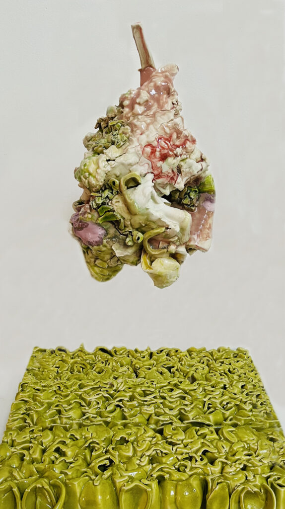
The two pieces created are linked by Dr. Coon’s research process as well as the progress of our meetings in developing the artwork.
Lost Memory in Matter relates to Dr. Coons discussions about Dementia. The abstracted form represents the mind and all that it can occupy. Dr. Coon gains access of information for an understanding of specific patient needs, from their initial fear to acceptance, with the goal being to balance the decline. I utilized layering as a way to present repetitive behavior modifications in order to achieve balance.
According to Dr. Coon, it is the perception of a shrinking world which can cause patients’ stress. However, building skills is a way to mold behavior and manage the situation while creating a safe space for dialogue. That space is established by the negative space in between the individual and their grounding of reality.
Behavioral Complexity is the framework where the response is equal to behavior and captures memories. Green is utilized to increase visual attention as it is the last color dementia patients loose the ability to see.
The Researcher: Dr. David Coon
Associate Dean of Research Initiatives, Support, and Engagement and Professor in the Edson College of Nursing and Health Innovation at ASU. He also serves as the director of the Center for Innovation in Healthy & Resilient Aging. Dr. Coon designs and evaluates interventions, such as CarePRO (Care Partners Reaching Out) and EPIC (Early-stage Partners in Care), that focus on culturally diverse groups of midlife and older adults facing chronic illnesses (e.g., dementia, cancer, depression) and their family caregivers.
The Artist: Susan Beiner
Beiner lives and works in Phoenix, AZ where she is Joan R. Lincoln Endowed Professor at Arizona State University and teaches courses in ceramics sculpture as well as art and science. Beiner’s art practice spans room size installations integrating landscape with environmental issues of ceramics and multi-media to achieve opulent effects.
Her ceramic work has been exhibited at The Mint Museum of Craft and Design, Limogues Foundation, Bernaudard France, Houston Center for Contemporary Craft, The Clayarch Gimhae Museum Korea, and the Princessehof Keramiekmuseum in the Netherlands to name a few. Beiner’s work is included in numerous collections including the The Los Angeles County Museum of Art, and National Museum of Ceramics in Leeuwarden, Netherlands, and the Yixing Ceramics Museum, China as well as other permanent and private collections. In 2020, she received the grand prize at the Taiwan Ceramics Biennale held at the Yingge Ceramics Museum, New Taipei City, Taiwan.
More Information
Lost Memory in Matter
Medium: 14” x 8” x 14” and 3” x 18” x 15”, porcelain with steel mount.
Retail cost: $3,000
Behavioral Complexity
Medium: 17” x 25” x 7.5”, porcelain, powder coated aluminum (Currently not completed and not pictured)
Retail cost: $3,000